Precise Time-Controlled Cryo-Optical Microscopy Advances

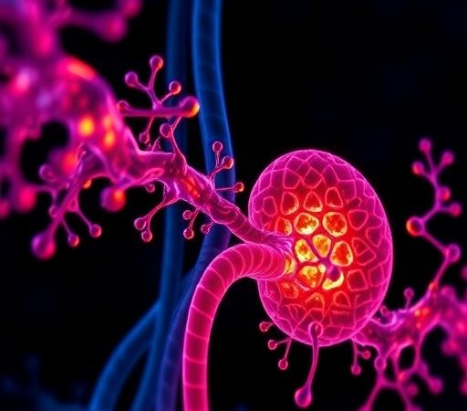
In a groundbreaking advance that promises to redefine the landscape of biological imaging, a team of scientists has unveiled a novel technique known as Time-Deterministic Cryo-Optical Microscopy. This innovative approach bridges the long-standing gap between temporal precision and cryogenic preservation, offering an unprecedented window into the dynamic molecular architecture of life at ultra-low temperatures. The method, detailed in the latest issue of Light: Science & Applications, heralds a new era in microscopy by enabling researchers to capture exquisitely timed snapshots of biological specimens with nanoscale resolution under cryogenic conditions, thereby preserving native biomolecular states while revealing dynamic processes that were previously inaccessible.
Traditional cryo-optical microscopy techniques have revolutionized structural biology by immobilizing samples in vitreous ice, thus maintaining their pristine native conformations. However, these methods have suffered from a critical limitation: the inability to precisely control and synchronize the timing of image acquisition relative to rapidly occurring biological events. This temporal uncertainty has posed a formidable challenge, particularly for studies aiming to elucidate transient states or fast kinetics at the molecular level. Addressing this, the team led by Tsuji, Yamanaka, Kumamoto, and colleagues has engineered an optical platform that integrates sophisticated timing control with cryogenic conditions, resulting in what they term “time-deterministic” imaging.
At the core of this breakthrough lies a custom-engineered cryostat system that couples ultra-fast optical shutters and pulsed excitation sources with cryo-temperature sample holding stages. This synergistic setup enables the precise triggering of illumination and detection windows with microsecond accuracy. Through meticulous synchronization of laser pulses with the sample’s cryogenic freezing and thawing cycles, the researchers can freeze biological activity at specific time points, capturing ultra-high-resolution images that faithfully reflect the structural state of biomolecules at those instants. This represents a quantum leap from prior methodologies, which typically recorded static or averaged images without temporal discrimination.
.adsslot_8iZlm2bYwR{width:728px !important;height:90px !important;}
@media(max-width:1199px){ .adsslot_8iZlm2bYwR{width:468px !important;height:60px !important;}
}
@media(max-width:767px){ .adsslot_8iZlm2bYwR{width:320px !important;height:50px !important;}
}
ADVERTISEMENT
The implications of time-deterministic cryo-optical microscopy extend far beyond mere technical innovation. By capturing biomolecular architectures at defined moments during dynamic processes—such as protein folding, enzymatic reactions, or conformational shifts—scientists can now explore the mechanistic underpinnings of life with both spatial and temporal acuity. For instance, the capacity to observe intermediate folding states of proteins frozen precisely as they occur sheds new light on diseases linked to protein misfolding. Similarly, enzyme catalysis, long a subject of static structural studies, can be interrogated through snapshots matched exactly to reaction intervals, revealing transient conformations central to biological function.
Implementing this system required overcoming several formidable engineering hurdles. Cryogenic microscopes are inherently sensitive to thermal fluctuations and mechanical vibrations, which can severely compromise image quality and temporal precision. The team expertly mitigated these issues by designing vibration-damped cryostats integrated with feedback-controlled temperature regulation. Additionally, optical components were optimized for minimal aberrations and maximal light throughput at very low temperatures. The use of custom-built pulsed laser systems with precisely controlled timing sequences ensured that excitation and emission signals corresponded exactly to the target temporal window. Collectively, these refinements coalesced into an imaging platform with spatial resolution at the single-nanometer scale and temporal timing with microsecond resolution.
Moreover, the researchers incorporated advanced image processing algorithms tailored to the unique noise characteristics of cryogenic optical data. Since ultra-low temperatures suppress thermal noise yet introduce other artifacts related to electronic sensors and photon counting, computational techniques were essential to enhance contrast, deconvolve signals, and extract meaningful structural information. This holistic approach, combining hardware precision with bespoke software, maximizes the fidelity of the resulting datasets, enabling confident interpretation of complex biological phenomena.
Among the key demonstrations showcased in the study, the team explored the structural dynamics of mitochondrial ATP synthase, a vital molecular motor responsible for cellular energy production. By applying time-deterministic cryo-optical microscopy, they captured sequential snapshots documenting conformational changes during different stages of ATP synthesis. These observations revealed hitherto unappreciated intermediate states that are crucial to understanding the enzyme’s efficiency and regulation. The ability to freeze and image these states on demand opens new avenues for drug discovery targeting metabolic disorders and mitochondrial dysfunctions.
The versatility of this approach is further underscored by its compatibility with diverse labeling strategies, including fluorescent protein markers, organic dyes, and quantum dots. This flexibility permits the selective highlighting of specific molecular components within complex assemblies, allowing multicolor and multimodal imaging under cryogenic conditions. Consequently, researchers can dissect spatial and temporal relationships among multiple biomolecules simultaneously, unraveling complex cellular machinery with deep molecular context.
From a broader perspective, time-deterministic cryo-optical microscopy offers transformative potential for fields spanning structural biology, biophysics, materials science, and nanotechnology. In addition to biological specimens, the technique can be adapted to study transient states of novel nanomaterials, polymers, and catalytic surfaces under cryogenic conditions, where dynamic processes occur on fast timescales yet require immobilization for optical interrogation. This cross-disciplinary applicability highlights the technology’s far-reaching impact.
Looking ahead, the research team envisions integration of this method with complementary cryo-electron microscopy (cryo-EM) and cryo-soft X-ray tomography techniques. Such correlative microscopy workflows would combine the unparalleled temporal control of time-deterministic cryo-optics with the elemental and ultrastructural resolution of electron and X-ray methods. This synergy could provide holistic snapshots of biological systems, resolving molecular structure, function, and dynamics seamlessly across multiple scales.
Another prospective development involves augmenting the temporal resolution further by employing ultrafast laser systems capable of femtosecond or even attosecond pulses. This would open the door to observing electron dynamics and chemical bond rearrangements in real time, under cryogenic preservation. Coupled with advances in computational microscopy and artificial intelligence-driven image analysis, these enhancements promise to accelerate discovery cycles and deepen our molecular understanding of life.
In the context of clinical research, time-deterministic cryo-optical microscopy may revolutionize pathological investigations by enabling snapshot imaging of disease-relevant molecular events from patient-derived samples. The ability to pinpoint structural and dynamic aberrations with high spatiotemporal resolution could facilitate early diagnosis, prognosis, and tailored therapeutic approaches for conditions including cancer, neurodegeneration, and infectious diseases.
The development of this technology also raises important questions about data management and storage, given the expected volume and complexity of time-resolved cryo-imaging datasets. The authors note ongoing efforts to establish robust computational infrastructures and standardized data formats to support collaborative analysis and reproducibility, ensuring that this powerful tool benefits the global scientific community.
In summary, the advent of time-deterministic cryo-optical microscopy represents a landmark achievement in optical microscopy, marrying cryogenic preservation with precise temporal control. By enabling researchers to freeze and image biological structures at exact moments during dynamic processes, this technique unveils molecular mechanisms with clarity and detail previously thought unattainable. As it integrates with existing methodologies and evolves further, it promises to catalyze revolutionary insights across disciplines, from fundamental biology to translational medicine and innovative materials science.
This pioneering research, authored by Tsuji, Yamanaka, Kumamoto, and their collaborators, illustrates the transformative power of interdisciplinary innovation and meticulous engineering. It sets a new benchmark for the exploration of life’s molecular dance, frozen yet alive in time, forever expanding the frontier of scientific imaging.
Article References:
Tsuji, K., Yamanaka, M., Kumamoto, Y. et al. Time-deterministic cryo-optical microscopy. Light Sci Appl 14, 275 (2025). https://doi.org/10.1038/s41377-025-01941-8
DOI: https://doi.org/10.1038/s41377-025-01941-8
Image Credits: AI Generated
Tags: biological imaging advancementsbridging cryogenics and opticscapturing transient molecular statescryogenic preservation techniquesdynamic molecular architecturefast kinetics imaging methodsimaging biological specimens at ultra-low temperaturesnanoscale resolution microscopynovel microscopy techniquesstructural biology innovationstemporal precision in microscopyTime-Deterministic Cryo-Optical Microscopy