Microstructured fiber measures the size of nanoparticles

by Stefanie Miethbauer, Leibniz-Institut für Photonische Technologien e. V.
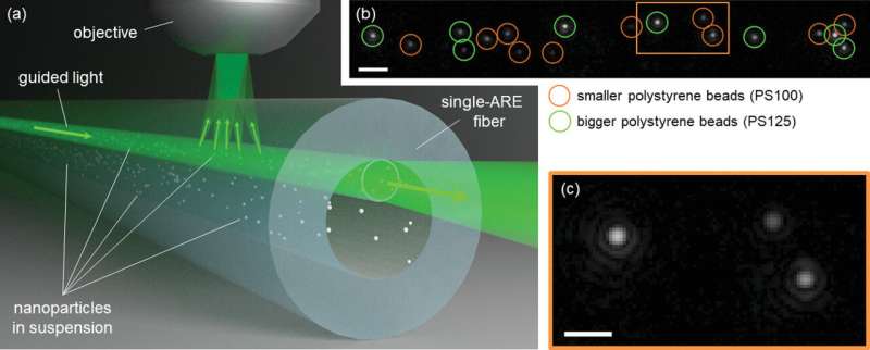
 2, 10 μm scale bar, gamma 0.4) with tracked nanoparticles from a bimodal mixture. Sample species: polystyrene (PS) beads with nominal diameters 100 nm (circled in orange) and 125 nm (circled in green). The full frame is included in Supporting Information IV. c) Image detail from (b) with visible first Airy rings confirming the aberration-free imaging quality (5 μm scale bar; more details on the image quality analysis can be found in Supporting Information IV). Credit: Small (2022). DOI: 10.1002/smll.202202024″ width=”800″ height=”323″>
Researchers at Leibniz Institute of Photonic Technology (Leibniz IPHT) developed a new glass fiber design that enables exceptionally long observations of a large number of individual, freely moving nanoparticles in a liquid. This allows the size distribution of nano-objects in a sample to be determined with even higher precision. The scientists are thus laying the foundation for even better research into environmental and bioanalytical issues in the future.
Whether water analysis, vaccine production or the examination of biological samples—mixtures of minute particles occur in almost all areas of everyday life and are composed of a variety of different tiny objects in liquid environments.
The accurate determination of individual components of such a fine particle mixture within a liquid (dispersion) poses challenges to science—especially with regard to the width of their size distribution and the presence of various particle species that differ only slightly in size. A new microstructured glass fiber (single antiresonant element fiber) developed at Leibniz IPHT offers the potential to significantly enhance the measurement accuracy of nano-object size characterization.
New optical fiber for high-precision analysis
With the special optical fiber realized at the Jena institute, nano-objects in aqueous solution with a diameter smaller than 20 nanometers can be confined, individually tracked and their size precisely determined. This allows researchers to precisely analyze size distributions of nanoparticles in mixtures. For this purpose, the glass fiber has a thin-walled and therefore light-conducting microchannel that’s 17 micrometers in diameter.
To examine a sample, the particle fluid is brought into contact with the hollow-core fiber, which fills with the fluid sample as a result of capillary force. The coupled light is guided along the fiber’s integrated fluid channel. The glass wall, which is only 756 nanometers thick, allows intense and uniform illumination of the sample to be examined and the contained nano-objects.
The light scattered by individual nanoparticles allows their position to be tracked and thus enables highly accurate microscopic observations. “With our new fiber-optical method, individual nanoscaled objects can be tracked over long periods of time. In this way, we can determine their size extremely accurately and reliably, so that we can characterize individual components in a mixture,” explains Mona Nissen, doctoral student in the Fiber Photonics Department at Leibniz IPHT.
In experimental studies with mixtures of particles with a small size difference, consisting of polystyrene nanospheres with average diameters of 100 and 125 nanometers, the researchers were able to demonstrate a high precision characterization using the novel optical fiber. The scientists were able to precisely measure the size distribution and identify individual components both in monodisperse particle mixtures with nano-objects of one species and size class and in polydisperse particle compositions with objects of different properties and sizes.
Nanoscale applications
The presented fiber-optical approach offers the potential to be used in nanotechnological applications in the field of environmental and bioanalytics as well as in chemistry and medicine for size control of nanoparticles. The researchers see application scenarios, for example, in the examination of waters for microplastic residues, the analysis of patient samples such as urine, the observation of synthesis products in chemical sciences or the development of drugs.
The findings are published in the journal Small.
More information:
Mona Nissen et al, Nanoparticle Tracking in Single‐Antiresonant‐Element Fiber for High‐Precision Size Distribution Analysis of Mono‐ and Polydisperse Samples, Small (2022). DOI: 10.1002/smll.202202024
Journal information:
Small
Provided by
Leibniz-Institut für Photonische Technologien e. V.
Citation:
Microstructured fiber measures the size of nanoparticles (2022, December 6)
retrieved 6 December 2022
from https://phys.org/news/2022-12-microstructured-fiber-size-nanoparticles.html
This document is subject to copyright. Apart from any fair dealing for the purpose of private study or research, no
part may be reproduced without the written permission. The content is provided for information purposes only.


